Cracked teeth — how they happen and how they're treated
A cracked tooth can be surprisingly hard to pin down — the discomfort comes and goes, and the crack itself is often invisible to the naked eye. Under the dental operating microscope I can find cracks early, work out how deep they run, and plan the right treatment to give the tooth its best chance of staying in function.
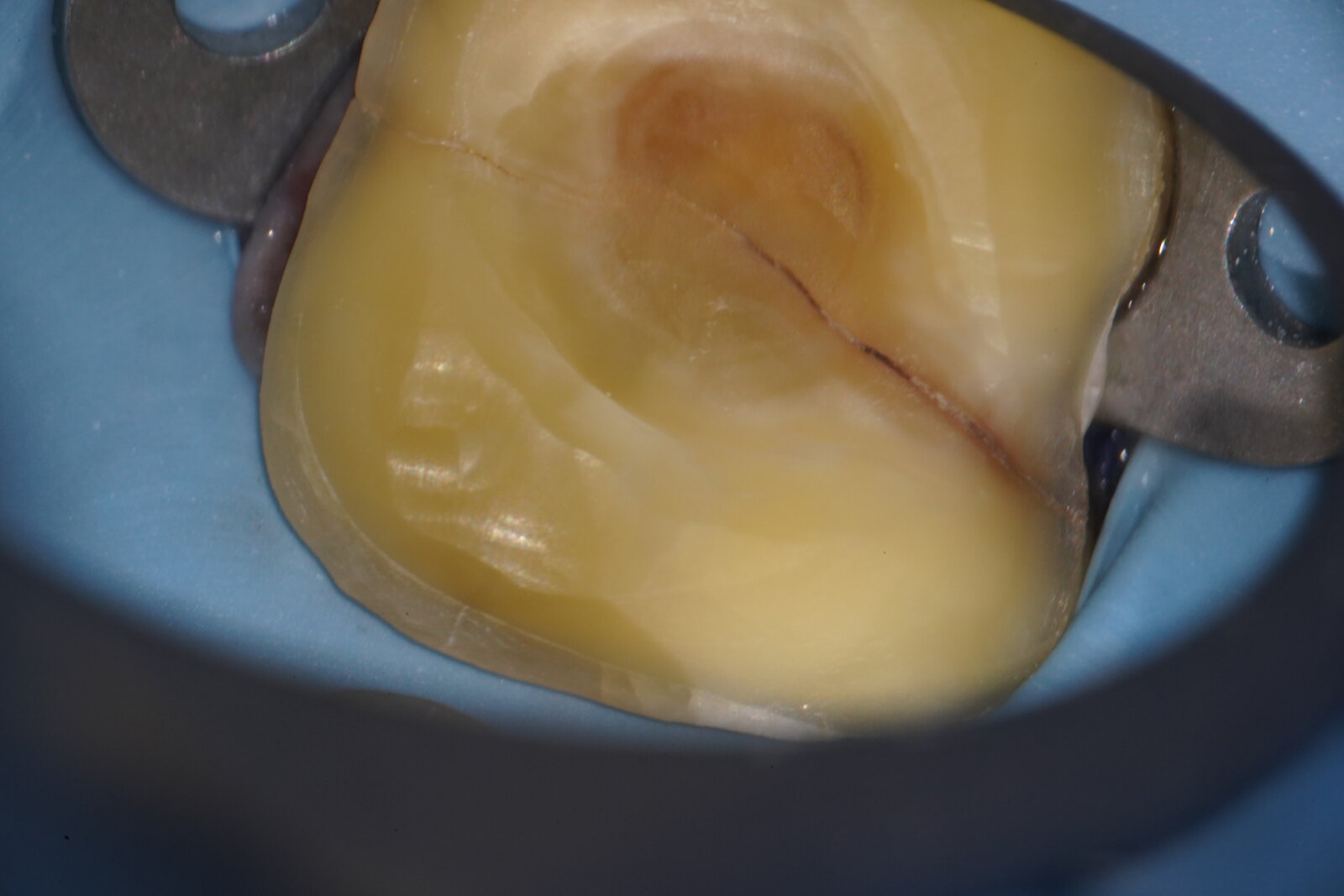
A cracked molar under the dental operating microscope — the fracture line runs right across the biting surface.
What is a cracked tooth?
A crack is a line of damage running through the hard structure of a tooth. Some are tiny lines in the surface enamel that never cause trouble; others run deeper, into the dentine or towards the nerve, and these are the ones that cause symptoms and need treating. Because a crack can open slightly under pressure and close again, it may be there for months or years before it's picked up — which is exactly why the discomfort tends to come and go.
Why teeth crack
Teeth crack for a few main reasons. The most common is everyday biting force — years of heavy chewing, and especially grinding or clenching (often overnight), place enormous repeated load on a tooth. A sudden injury or knock can crack a tooth outright. And teeth that already carry large fillings are more vulnerable, because less natural tooth remains to absorb the load — this is especially true of older amalgam (silver) fillings, which don't bond to the tooth and can act like a wedge over many years.
Signs you might have a crack
The classic sign is a sharp pain when you bite down or, tellingly, when you release the bite. You might notice sensitivity to cold or sweet things, or an ache you can't quite place to a single tooth. Symptoms often come and go, which can make a crack frustrating to track down — but that on-and-off pattern is itself a useful clue that points towards a crack.
Finding the crack
Cracks are often invisible to the naked eye, so I examine the tooth under the dental operating microscope, where fine lines stand out under strong, magnified light. Shining light through the tooth, and where helpful a dye that seeps into the crack, can reveal its path. Sometimes the clearest picture only appears once an old filling is removed and the crack can be traced across the tooth.
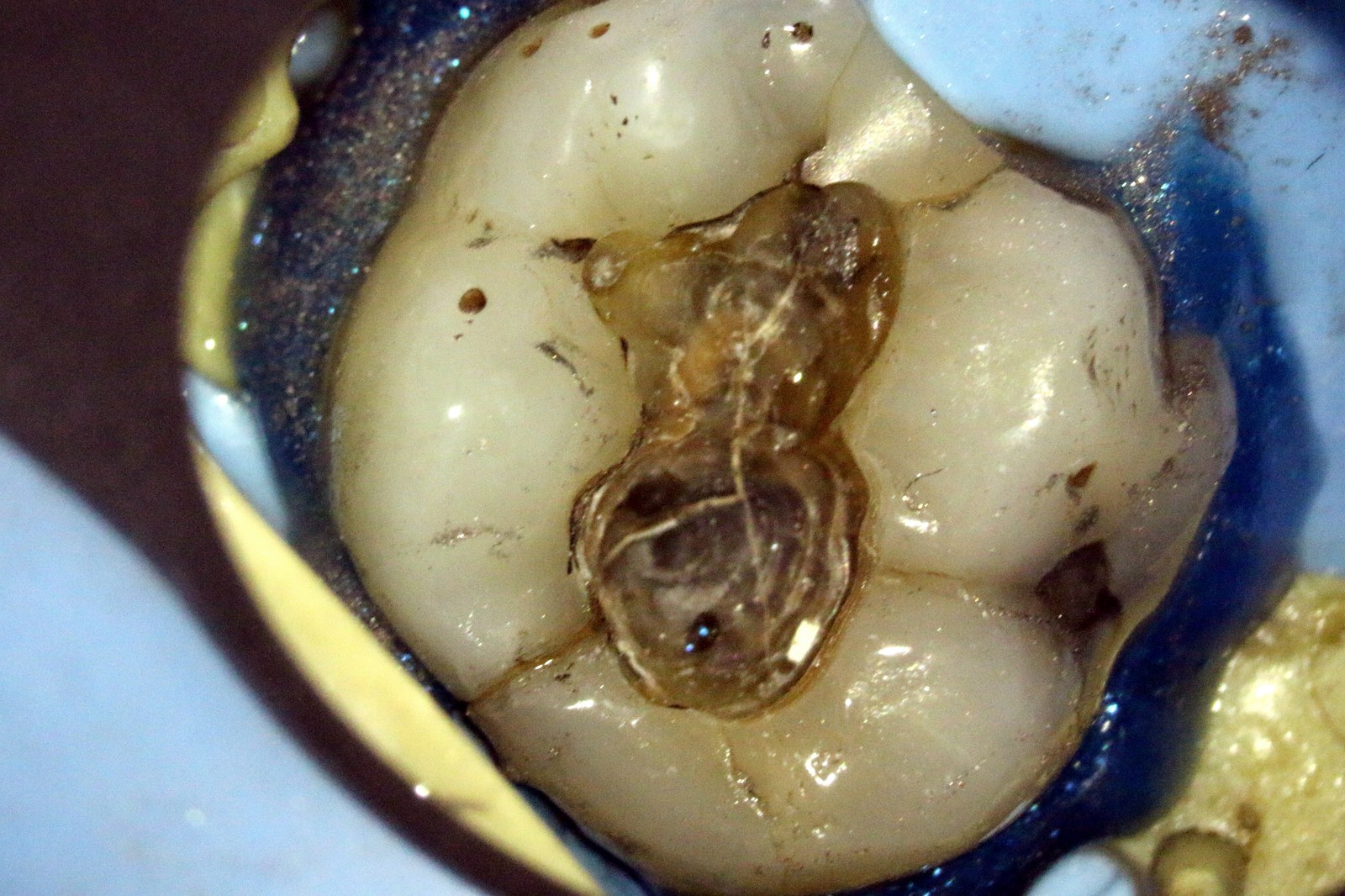
Under the microscope, once an old filling is removed, the crack can be traced across the tooth.
How a cracked tooth is treated
Treatment depends on how deep the crack runs. Caught early, a crack can often be managed by removing the weakened, cracked structure and rebuilding the tooth so that biting force is taken off the crack. If the crack has reached the nerve and caused lasting damage, root canal treatment may be needed to settle the tooth and keep it. Occasionally a crack runs too far — down into the root — and the tooth can't be saved; finding that out early, and honestly, matters just as much. Where grinding or clenching is part of the picture, a well-made nightguard helps protect the tooth going forward.
A cracked molar restored under the dental operating microscope — the crack, isolating and building the tooth back up, and the finished result.
Working together to restore your tooth
Saving a cracked tooth is often a two-part job. There's the endodontic side — assessing how far the crack runs and, where the nerve is involved, carrying out root canal treatment to settle the tooth — and the restorative side, rebuilding it so it can take full biting force again, usually with a cusp-covering onlay or crown that holds the tooth together and stops the crack spreading.
I work closely alongside expert restorative dentists, so both sides are planned and carried out as one joined-up treatment rather than a series of disconnected appointments. You're looked after by a team that's already talking to each other, with a single shared goal: getting your own natural tooth back to comfortable, lasting function.
At Bellevue, Sardinia House and Widegate House, we also have the technology to design and fit a crown in a single day. The tooth is scanned digitally and the crown made and placed in the same visit, so there's no temporary crown, no second appointment and no weeks of waiting between stages — you leave with the tooth fully restored, protected and back in normal function.
Get in touch